Syngene is where innovation meets excellence in life sciences, molecular biology, genomics, and proteomics. As a division of Synoptics Ltd, Syngene is committed to pushing the boundaries of scientific research through our image analysis technology. From gel documentation to fluorescence and chemiluminescence imaging equipment, our products are meticulously crafted to meet the rigorous standards set by regulatory agencies and accreditation bodies. Cameras are pivotal in our mission, underscoring our dedication to delivering unparalleled quality and performance.
Western blot imaging: Which CCD camera should I choose for maximum sensitivity?
Whether using chemiluminescence or fluorescence, Western blot is the most popular protein detection method when studying proteins. There are two commonly used methods for detecting chemiluminescence signals:
- Exposing the western blot to X-ray film.
- Use a digital imaging system with a cooled charged coupled device (CCD) camera.
CCD cameras employ a light-sensitive silicon chip to transform photons into digital signals. Cutting-edge systems, like the G:Box chemi range, offer digital imaging capabilities that outperform film detection methods. These advancements include heightened sensitivity, improved linearity, and expanded dynamic range, facilitating accurate protein quantification across a wide range of concentrations spanning four orders of magnitude (4OD).
What should you consider when choosing a CCD camera?
One of the primary considerations is how sensitive the CCD camera is. When assessing the sensitivity of the camera, you also need to look at some key specifications, such as:
- Signal-to-noise ratio (SNR)
- Quantum efficiency (QE) percentage
- Pixel size
- Dynamic range
Understanding these key specifications will allow you to evaluate the imaging system that suits your needs.
Signal-to-noise (SNR) ratio
A researcher performing Western blots aims to maximise the signal from the protein band while minimising the membrane’s background. This is known as the signal-to-noise ratio (SNR). To achieve the best SNR ratio, you need to capture as much of the signal as possible (highest quantum efficiency percentage) and reduce the sources of noise to a minimum.
I will explain quantum efficiency more later, but to understand the SNR ratio, you need to know the different types of noise generated by the CCD sensor. One main source of noise is known as readout noise. So, what is readout noise? It is a combination of noise sources originating from the process of amplifying and converting the photoelectron to a voltage. The faster the camera read out, the higher the noise can be.
Dark noise is the second noise source, arising from heat generated without any signal applied. Cooling the camera reduces the dark signal and permits longer integration periods, up to several hours. For chemiluminescent imaging without significant dark noise accumulation, it is essential to check that you use a cooled camera. All the G:Box chemi range cameras are Peltier cooled, ensuring the dark noise is kept to a minimum when capturing images with a long exposure time.
Incoming photons have an inherent noise known as photon shot noise. If you measured the signal from a calibrated and constant light source, the value would fluctuate and appear to be noise from the camera. However, this effect can be attributed to the variability of the signal itself.
Quantum Efficiency (QE)
Quantum Efficiency (QE) is the sensor’s ability to convert incoming photons into a measurable electronic signal. QE is often expressed as a percentage; therefore, a QE of 80% indicates an 80% chance that a photoelectron will be released for each incident photon.
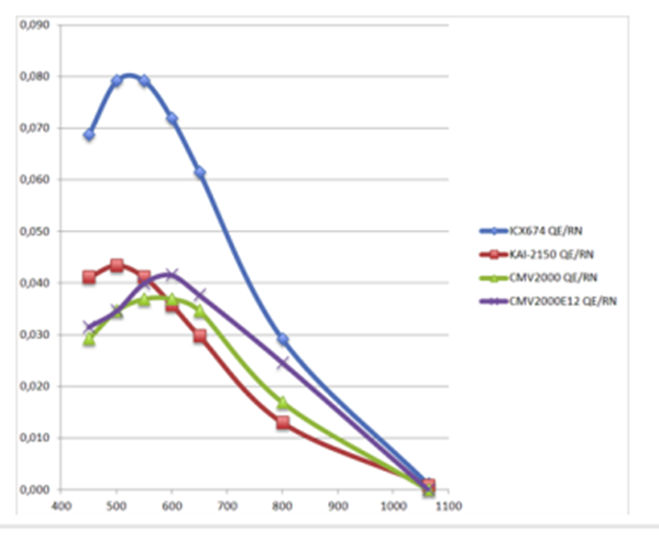
QE is wavelength dependent (photon energy), and therefore, it is important to check the QE curve of the camera sensor at the wavelength of interest. The G:Box Chemi range cameras use the sensor ICX674 and have a high QE of 73% at 425nm, which makes these systems ideal for capturing chemiluminescence Western blots as chemiluminescence emits at a wavelength of around 425nm (refer to graph). When imaging fluorescence, it is essential to check the QE curve at the wavelength of interest for the fluorescent dye you are imaging to ensure you will achieve maximum sensitivity from the sensor camera.
Pixel Size
A pixel in a camera sensor collects photons of light and converts them into an electrical signal. The signals are digitised, and the camera can generate the image from the values received at each pixel.
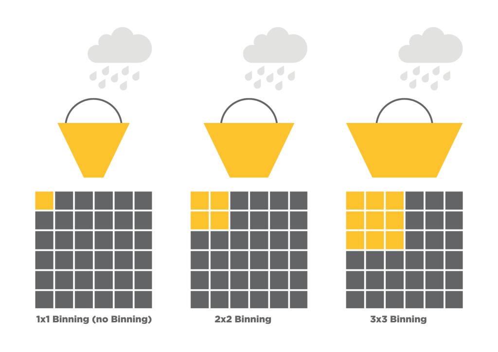
Smaller pixels can be binned, which means that the signal in adjacent pixels on a sensor can be combined to create a single larger effective pixel (known as a superpixel), increasing sensitivity and, therefore, shortening exposure times. The downside of binning is that it does lead to a loss in resolution. The 3 bucket graphic explains this with rain being captured in buckets. The larger buckets can capture a bigger volume of raindrops; therefore, larger pixels can capture bigger photons.
The demand for high-megapixel sensors has led to developments in sensor design and enabled ever-smaller pixels to be packed into sensors. While having more pixels can be beneficial, there are tradeoffs, as pixel size can impact signal-to-noise and dynamic range. Therefore, it is important to consider your application when choosing the pixel size for your gel doc.
Dynamic Range
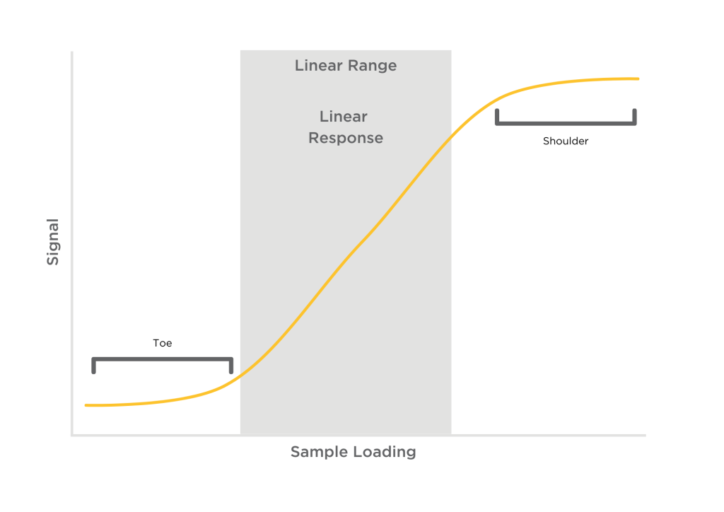
The dynamic range of a CCD camera is the range the camera can capture in a single image, a linear response to a signal from the weakest to the strongest (see line graph).
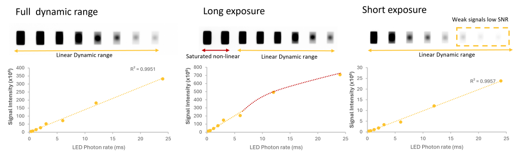
Many researchers desire to analyse both low- and high-expressing proteins in the same Western blot sample. Imaging these two things simultaneously can be challenging for imaging systems with a limited dynamic range. Capturing images with long exposure times permits the detection of low-expressing proteins and results in saturated or ‘blown out’ bands.
Systems with a wide dynamic range allow image capture of both the low and high-intensity bands, permitting the analysis of weakly and strongly expressed proteins.
Understanding dynamic range is essential for properly analysing your data and interpreting your Western blotting results. Accurate quantification requires the signal from protein bands to be within the linear range of the imaging system. The relationship between band intensity and protein quantity within the linear range is linear and proportional. Signals outside the linear range can be affected by noise if they are weak or too strong, and then the imaging system can no longer accurately record the increasing signal (saturation), as shown in the photo.
When looking at the dynamic range of a CCD camera, you also need to consider the full capacity of your sensor. Why is it important to know the full-well capacity? Full-well capacity is the largest charge a pixel can hold before saturation, which results in signal degradation. When the charge in a pixel exceeds the saturation level, the charge starts to fill adjacent pixels, a process known as blooming, resulting in image smearing. When the pixel is saturating (blooming), the linear response of the CCD will begin to deviate and compromise the quantitative performance.

In conclusion, a camera’s sensitivity is affected by several factors. When determining the sensitivity of a CCD camera, take a close look at the specifications:
- dark noise
- readout noise
- the quantum efficiency percentage
- the pixel size
- dynamic range
- full-well capacity
Check out the G:Box chemi range and the new G:Box mini XRQ!